Dr Bhavendu D. L, Thiruvananthapuram

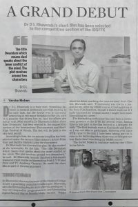
New India Express, 25th May 2017
Dr: Bhavendu, Memories of IFFK | IFFK News


Interview by Dr. Maggie Xavier with Dr D L Bhavendhu—a passionate filmmaker.


Q: Sir can you tell us briefly about yourself?

A: I am  from Thiruvananthapuram

I did my MBBS and DMRD and DNB from Thiruvananthapuram medical College, Worked as adhoc Lecturer in Medical college hospital, Thiruvananthapuram. [ 2005—2012]. Now working as Cons; Radiologist  at Ananthapuri Hospitals Thiruvananthapuram past 6 yrs

Q: In which modalities are you interested in?

A: I work in all modalities

Q: What are your areas of special interest?

A: Passionate about world cinema. Hobbies are photography, music and reading.

Q: Can you say something about your achievements?

A: First short film got selection in the competition section of the International Documentary And Short Film Festival Of Kerala 2017. I am a film society member

Q: Can you tell us about your life as filmmaker?

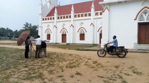
A: Before two years I went for a course in film making. There is a film academy in Chennai, which has a branch in Thiruvananthapuram. I went for a course there. It was on weekends. We are given training in pre-production, production and post-production. At the end of the course, every batch of students has to do a film. I chose a Malayalam short story, based on which I did my film.

Q: Who all help you? Or who are the team members?

A: The academy helps us. My batchmates also help me. We work as a group, but the chunk of the work is done by me. I chose the actors and actresses.Mrs.Sajitha Madathil has acted in my film.

Q: Where did you shoot? Budget?

A: Shooting was mostly in Thiruvananthapuram. We have to bear all the expenses. The academy will help us a bit.

Q: What Was The Theme

A: I chose a Malayalam short story, based on which I did my film. This is my first short film. The film was named “DWANDHWAM”. This six-minute long film titled DWANDHWAM means DUEL which speaks about the inner conflict of mind. The plot revolves around two characters.

Q: Sir I heard you are on to your second short film. Can you tell us about it?

A: Yes. I also did a second short film DATHAM. Just got the mail that it has been accepted for the short film section of this year’s Cannes Film Festival.

Q: Sir can you tell us about your family?

A: Wife HSS Teacher, Son 12 Yrs, Daughter 9 Yrs

Journal Club: Magnetic resonance imaging spectrum of perinatal hypoxic ischemic brain injury

Sharing an article published by Dr Binoj Varghese, et al; that was awarded the BEST RESEARCH PAPER published in IJRI 2018, at the National IRIA conference at Chandigarh 2019


1. Indian J Radiol Imaging. 2016 Jul-Sep;26(3):316-327. Magnetic resonance imaging spectrum of perinatal hypoxic-ischemic brain injury. Varghese B, Xavier R, Manoj VC, Aneesh MK, Priya PS, Kumar A, Sreenivasan VK.

Erratum in Indian J Radiol Imaging. 2016 Oct-Dec;26(4):530.

Perinatal hypoxic-ischemic brain injury results in neonatal hypoxic-ischemic encephalopathy and serious long-term neurodevelopmental sequelae. Magnetic resonance imaging (MRI) of the brain is an ideal and safe imaging modality for suspected hypoxic-ischemic injury. The pattern of injury depends on brain maturity at the time of insult, severity of hypotension, and duration of insult. Time of imaging after the insult influences the imaging findings. Mild to moderate hypoperfusion results in germinal matrix hemorrhages and periventricular leukomalacia in preterm neonates and parasagittal watershed territory infarcts in full-term neonates. Severe insult preferentially damages the deep gray matter in both term and preterm infants. However, associated frequent perirolandic injury is seen in term neonates. MRI is useful in establishing the clinical diagnosis, assessing the severity of injury, and thereby prognosticating the outcome. Familiarity with imaging spectrum and insight into factors affecting the injury will enlighten the radiologist to provide an appropriate diagnosis.

DOI: 10.4103/0971-3026.190421 PMCID: PMC5036328 PMID: 27857456

Read complete article:

https://www.ncbi.nlm.nih.gov/pmc/articles/PMC5036328/

Journal Club: High prevalence of cervical perineural cysts on cervical spine MRI-Case series

CITATION: Gossner J. High prevalence of cervical perineural cysts on cervical spine MRI. Int J Anat Var. 2018;11(1): 18-19.

ABSTRACT
The Cervical perineural (Tarlov) cysts have been reported to be a rare occurance. The frequency, distribution and possible clinical relevance of such perineural cyst in the cervical spine were studied. In a retrospective review of
41 patients undergoing cervical spine MRI 6 patients showed perineural cysts (14%). Size ranged from 4 to 12 mm and the lower cervical was predominantly affected. The reported clinical symptoms could not be correlated to the found perineural cysts. Cervical perineural cysts seem to be a common asymtomatic incidental finding. The rare diagnosis of a symptomatic perineural cyst should be made with caution and is a diagnosis of exclusion.


READ THE COMPLETE ARTICLE: high-prevalence-of-cervical-perineural-cysts-on-cervical-spine-mri

Journal Club: From Inguinal Hernias to Spermatic Cord Lipomas: Pearls, Pitfalls, and Mimics of Abdominal and Pelvic Hernias

Citation: Miguel C. Cabarrus, MD, Benjamin M. Yeh, MD, Andrew S. Phelps, MD, Jao J. Ou, MD, PhD, Spencer C. Behr, MD. RadioGraphics 2017; 37:2063–2082.  https://doi.org/10.1148/rg.2017170070

Abdominal and pelvic hernias may be indolent and detected incidentally, manifest acutely with pain and distress, or cause chronic discomfort. Physical examination findings are often ambiguous and insufficient for optimal triage. Therefore, accurate anatomic delineation and identification of complications are critical for effective treatment planning. Imaging, particularly computed tomography, provides a vital understanding of the hernia’s location and size, involved viscera, and severity of associated complications. Reader familiarity with the imaging appearances and anatomic landmarks of hernias is important for correct diagnosis, which may impact preoperative planning and reduce morbidity. This article reviews the appearance of anatomic structures in the abdominal wall and pelvis that are important for diagnosing common and uncommon abdominal and pelvic hernias, and it highlights key imaging features that are helpful for differentiating hernias, mimics, and their complications.

Read the Complete Article